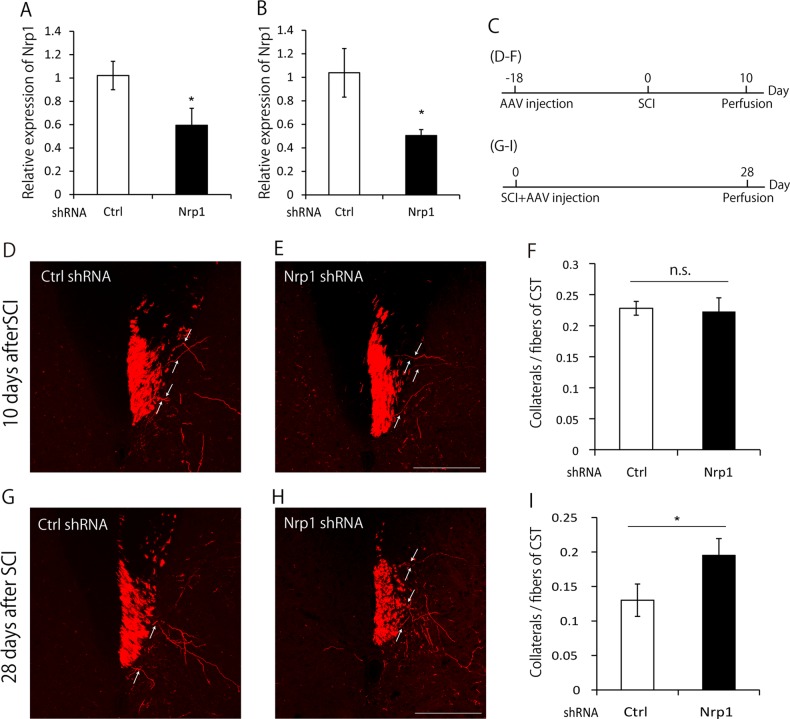
Fig. 3. Neuropilin-1 (Nrp1) knockdown suppresses the pruning of collaterals.
a Quantification of Nrp1 mRNA by real-time PCR analysis in DIV4 cultured E16 mouse cortical neurons transfected with control shRNA or Nrp1 shRNA. The expression of Nrp1 was significantly suppressed by Nrp1 shRNA. b Quantification of Nrp1 mRNA by real-time PCR analysis in the motor cortex dissected by laser microdissection at 28 days after the AAV injection. The expression of Nrp1 was significantly suppressed by AAV expressing Nrp1 shRNA compared to that expressing control shRNA. c Experimental time course. To visualize the collaterals at 10 days after spinal cord injury (SCI), AAV was injected into the hindlimb motor area at 18 days before SCI. Then, 10 days after SCI, mice were perfused. To visualize the collaterals at 28 days after SCI, AAV was injected immediately following SCI, and mice were perfused at 28 days after SCI. d, e Transverse cervical sections showing turbo red fluorescent protein (tRFP)-labeled corticospinal tract (CST) axons (red) in mice infected with AAV expressing control shRNA (d) or Nrp1 shRNA (e) at 10 days after SCI. White arrows indicate collaterals sprouted from CST. Scale bar: 200 μm. f Quantification of the number of collaterals at 10 days after SCI. The number of collaterals in Nrp1 knockdown mice and control mice did not show the significant difference. g, h Transverse cervical sections showing tRFP-labeled CST axons (red) in mice infected with AAV expressing control shRNA (g) or Nrp1 shRNA (h) at 28 days after SCI. White arrows indicate collaterals sprouted from CST. Scale bar: 200 μm. i Quantification of the number of collaterals at 28 days after SCI. The number of collaterals was higher in Nrp1 knockdown mice compared with control mice. Data are presented as mean ± SEM. n = 4 (A), 3 (B), 6–8 (F), 6 (I), *p < 0.05; n.s. no statistical significance, Student’s t-test

