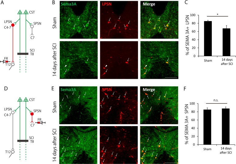
Fig. 5. Semaphorin 3A (Sema3A) expression is decreased in long propriospinal neurons (LPSNs) 14 days after spinal cord injury (SCI).
a Scheme of the experiment in (b) and (c). To label LPSNs, the retrograde tracer, Fluoro-Ruby (FR), was injected into T12. Immunohistochemistry for Sema3A was then performed. b Transverse cervical sections (C4-C7) showing Sema3A and FR-labeled LPSNs in sham (upper) and injured (lower) mice. White arrows indicate LPSNs expressing Sema3A. White arrowheads indicate LPSNs not expressing Sema3A. Scale bar: 200 μm. c Quantification of the ratio of LPSNs expressing Sema3A in cervical enlargement. The ratio was decreased after SCI. d Scheme of the experiment in (e) and (f). To label short propriospinal neurons (SPSNs), FR was injected into C7 level spinal cord. e Transverse cervical sections (C4-C7) showing Sema3A and FR-labeled SPSNs in sham (upper) and injured (lower) mice. Scale bar: 200 μm. f Quantification of the ratio of SPSN expressing Sema3A in cervical enlargement. Data are presented as mean ± SEM. n = 7–8 (c), 6–7 (f), *p < 0.05; n.s. no statistical significance, Welch’st-test (c), Student’s t-test (f)

