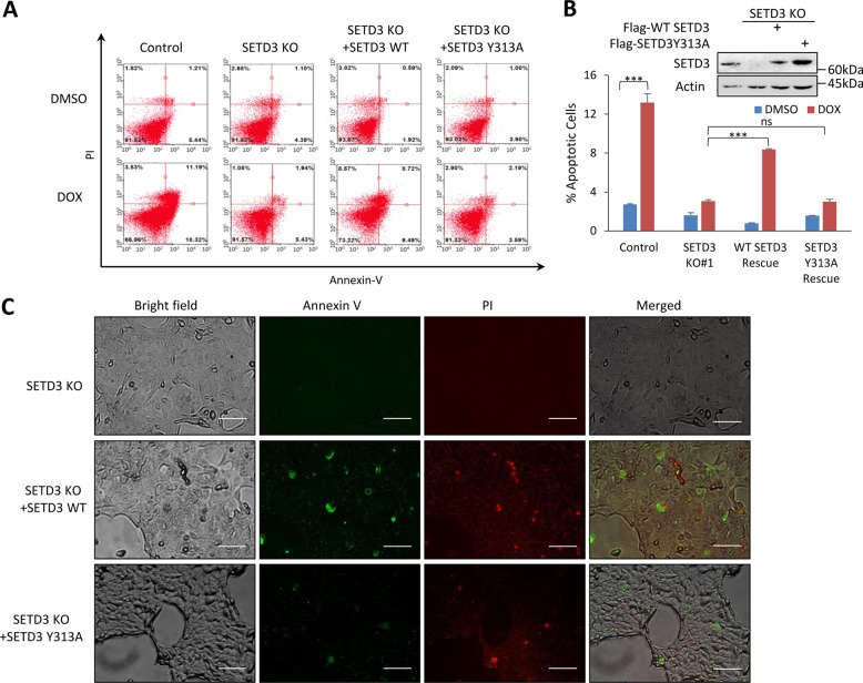
Fig. 3. DNA-damage-induced apoptosis is SETD3 and methylation dependent.
a FACS analysis of control, KO, WT SETD3 rescue and catalytic inactive (Y313A) SETD3 rescue cells, post DOX or vehicle treatment. b Quantification of apoptotic cells percentage of three-independent FACS analyses, ***p ≤ 0.001, ns p > 0.05. Western blot of the control, KO and rescued cells lysate. c SETD3 KO and rescued (with WT or Y313A) cells were stained with FITC-Annexin V and PI post treatment with DOX and viewed under fluorescent microscope (scale bar signifies 100 µM, pictures were taken under ×20 magnification)

