Fig. 2.
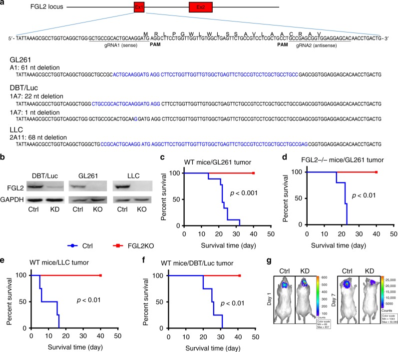
FGL2 knockout in tumor cells abolishes tumor progression. a Results of DNA fragment deletion in FGL2 exon 1 in individual clones were validated by gene sequencing. Paired gRNAs were designed to excise exon 1 at the mouse FGL2 locus. Individual clones isolated from cells transfected with gRNAs were assayed for deletions and inversions by RT-PCR. b Expression level of FGL2 in three tumor-cell lines, control (Ctrl) and FGL2-knockout (KO) or knockdown (KD) tumor cells, was detected by western blotting. The western blots shown represent three independent experiments. c Survival curve of wild-type (WT) immunocompetent C57BL/6 mice implanted with GL261-Ctrl or GL261-FGL2KO tumor cells (5 × 10⁴ cells per mouse; n = 9 per group). d Survival curve of FGL2−/− mice implanted with GL261-Ctrl or GL261-FGL2KO tumor cells (5 × 10⁴ cells per mouse; n = 5/group). e Survival curve of WT immunocompetent C57BL/6 mice implanted with Lewis lung cancer (LLC)-Ctrl or LLC-FGL2KO tumor cells (5 × 10⁴ cells per mouse; n = 4 per group). f Survival curve of WT immunocompetent BALB/C mice implanted with mouse astrocytoma (DBT)-Ctrl cells or DBT-FGL2KD tumor cells (5 × 10⁴ cells per mouse; n = 4 per group). g Bioluminescence imaging showing DBT-Ctrl and DBT-FGL2KD tumors at day 1 and day 7 after tumor-cell implantation. All data are representative of at least two independent experiments. The survival curves were analyzed by Kaplan–Meier analysis and the log-rank test was used to compare overall survival between groups

