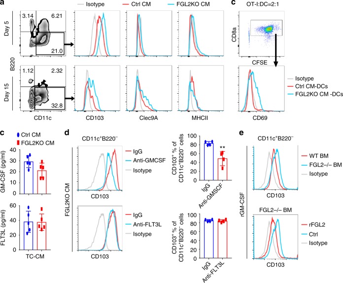
Fig. 5.
FGL2 suppresses GM-CSF-dependent CD103 induction on cDCs in vitro. a Expression of CD103 on CD11c+B220− DCs cultured with conditioned medium (CM) for 5 or 15 days. Bone marrow cells were isolated from C57BL/6 mice and then cultured with Ctrl-CM (CM from Ctrl tumor cells) or FGL2KO-CM (CM from FGL2KO tumor cells) for 5 days or 15 days. Non-adherent cells were recovered, stained, and analyzed by FACS to determine the expression levels of CD103, Clec9A, and MHCII on the CD11c+B220− DCs. b OT-I T cell activation by OVA-pulsed CM-cultured bone marrow DCs. Non-adherent bone marrow DCs were recovered from CM-cultured bone marrow DCs at 15 days. The bone marrow DCs (2 × 10⁴) were incubated with soluble OVA protein (100 μg/mL) for 2 h and then washed twice before co-culturing with the CFSE-labeled OT-I cells (5 × 10⁴). OT-I activation was measured by CD69 expression level, which was determined by flow cytometry. c Protein levels of GM-CSF and FLT3L in medium conditioned by tumor cells were detected by ELISA. Data were summarized from five independent experiments and were analyzed by paired t-test. d Representative FACS assay of CD103 expression on CD11c+B220− DCs cultured with FGL2KO-CM in the presence of neutralization antibody anti-GM-CSF (5 μg/mL) or anti-FLT3L (5 μg/mL) for 5 days. Data were summarized from four independent experiments and were analyzed by paired t-test. **P < 0.01 comparing with IgG. e Representative FACS assay of CD103 expression on wild-type (WT) and FGL2KO bone marrow (BM) cells cultured with rmGM-CSF (10 ng/mL) for 7 days with or without rFGL2 (200 ng/mL). All plots are representative of three independent experiments

