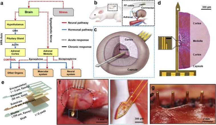
Fig. 1.
Schematic of the implanted device on the adrenal gland of a rat. (A) The scheme describing the stress response mechanism. When the brain recognizes the stress situation, neural and hormonal signals are transmitted from the brain to the lower organs, which represent acute and chronic responses, respectively. The adrenal medulla and adrenal cortex receive neural and hormonal signal, respectively, and perform the acute and chronic responses to stress. (B) The device is implanted in the dorsal part of the abdominal cavity (Left). The probe and the connector are linked with a conventional FFC (Right). (C) Detailed schematics of the sectional view of the adrenal gland and implanted probe. (D) The image of the probe taken by the optical microscope, and the relative sectional view of the adrenal gland. The four electrodes each have 700 μm of intervals (window size; 10 μm × 10 μm), so that they are able to cover both the cortex and medulla. (Scale bar in Inset: 50 μm.) (E) Schematic describing the structural information of the probe. (F) Photo image of the probe (yellow guideline) penetrating the adrenal gland (black dotted circle). The arrowhead tip fully penetrates the adrenal gland. (G) Photo image of the pins of the connector after implantation. Pin-based connection minimizes the possibilities of inflammation, and enables long-term recording.

