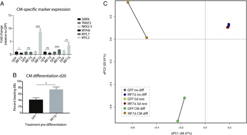
Fig. 6.
IRF7Δ dysregulates gene expression during cardiomyocyte differentiation. (A) qRT-PCR analysis of cardiomyocyte (CM)-specific markers from GFP- or IRF7Δ- expressing iPSC clones treated with Dox for 48 h before a 5-d rest. Levels of SIRPA, TNNT2, NKX2-5, MYH6, MYL7, and MYL2 mRNA are depicted as fold change over GFP. Error bars depict the SD of the mean. Significance was determined by unpaired Student’s t test where * to **** denote P values of 0.05 to <0.0001, respectively. (B) Graph quantifying the beating behavior of GFP- vs. IRF7Δ-pulsed iPSCs on day 20. (C) Analysis of the transcriptional variation between duplicate samples of untreated iPSCs, Dox-treated iPSCs, and cardiomyocyte differentiations for GFP and IRF7Δ lines.

