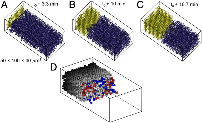
Fig. 2.
Growth of a BCC crystal from the liquid. (A–C) Reconstructed images of the crystallization process, where yellow particles are crystalline and blue particles are liquid. (D) Identification over one imaging interval of 4 s of attachment (red) and detachment (blue) sites at the crystal boundary with the liquid. The interface particles have been removed for clarity.

