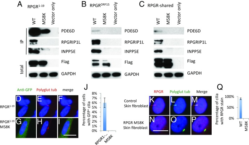
Fig. 5.
An M58K variation found in RP patients disrupts the interaction of RPGR with its interactors and ciliary localization of RPGR1−19. Flag-S-tag–conjugated WT and mutant RPGR isoforms and RPGR-shared region were transfected into HEK293T cells, and RPGR was pulled down by Flag beads. Western blots were used to detect RPGR interacting proteins. M58K variation disrupts the interaction between RPGR1−19 (A), RPGRORF15 (B), and RPGR-shared region (C) with endogenous PDE6D, INPP5E, and RPGRIP1L. GFP-tagged WT (D–F) and M58K (G–I) mutated RPGR1−19 transfected RPE1 cells show that M58K variation disrupts the RPGR1−19 ciliary localization. (J) Quantification of percentage of cells with GFP+ cilia in transfected RPE1 cells. Skin fibroblast cells from a patient with the M58K mutation lack RPGR cilia localization (N–P) compared with control skin fibroblast cells (K–M). (Q) Quantification of RPGR frequency of skin fibroblast cells. Each transfection was done three times. (Scale bars: 10 μm.)

