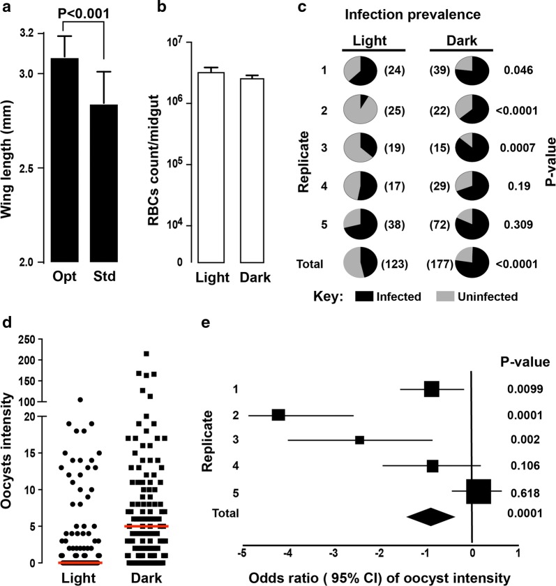
Fig. 2.
Mosquito infection with Plasmodium falciparum using the streamlined SMFA and mosquito dark-feeding and resting regime. a Comparison of wing length as a proxy for body size between mosquitoes reared as reported in this manuscript (optimal; opt) and mosquitoes generated under standard colony maintaining regime (std). b Comparison of RBC count in the mosquito gut bolus, used as a proxy to bloodmeal volume, between mosquitoes maintained in dark conditions for 3 h prior to, during and several hours after blood feeding (dark-feeding regime) compared mosquitoes fed using the standard SMFA protocol. c Pie-charts showing P. falciparum oocyst infection prevalence in mosquitoes under dark-feeding regime compared to mosquitoes fed using the standard SMFA protocol. Numbers in brackets show the number of mosquitoes used for each replicate as well as the total number of mosquitoes. d Overall P. falciparum infection intensities between mosquitoes under dark-feeding regime and mosquitoes fed using the standard SMFA protocol. e Forest plot showing estimate of odds ratio (± 95% CI) of oocyst intensities between mosquitoes under a dark-feeding regime and control. Squares and diamond shows the sample size for each replicate and the total, respectively

