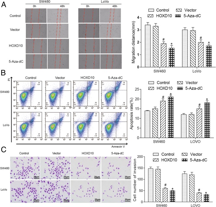
Fig. 7.
HOXD10 re-expression inhibited cell migration, invasion and promoted the cell apoptosis. a HOXD10 reduced the migration rates of SW480 and LoVo cells in scratch wound-healing assay, and photographs were taken at 0, 48 h after the wound was made (left). Statistical plot of the average number of migrated SW480 and LoVo cells in each group (right). * indicated P < 0.05 in comparison with the control group and # indicated P < 0.05 in comparison with the vector control group. b The result of flow cytometry showed HOXD10 overexpression induced SW480 and LoVo cells apoptosis (left). Statistical plot displayed percentages of apoptosis in SW480 and LoVo cells (right), *P < 0.05;#P < 0.05. c Overexpression of HOXD10 significantly decreased the invasive potential of both SW480 and LoVo cell lines through Matrigel invasion Transwell assay (left). Statistical plot of the average number of invaded SW480 and LoVo cells in each group (right). The graph showed the mean ± SD. *P < 0.05; #P < 0.05

