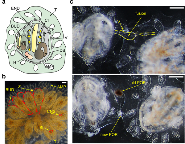
Figure 1. B. schlosseri Anatomy and Natural Transplantation Reactions.

a, Diagram of a zooid (ventral view) and primary bud (BUD), embedded within a tunic (TUN), with vasculature (V) connected to the zooid and bud which terminates in ampullae (AMP), the zooid has a branchial sac consisting of the endostyle (END) and stigmata (S), cell islands (CI), digestive system (DS) and heart (H). b, Live imaging of a colony (dorsal view), developing buds (BUD) are connected to the parental zooids (Z), all are connected to blood vessels, zooid’s siphons (SI) and central nervous system (CNS) are observed c, Live imaging of colonies undergoing fusion (top) and rejection (bottom), arrows point to fused vasculature and points of rejection (POR). Scale bar 0.2 mm.
