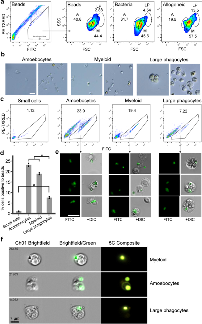
Extended Data Figure 7. Discovery of a Myeloid Lineage Phagocytic Population.

a, FACS analysis of B. schlosseri cells that are fluorescently positive in one of three phagocytosis assays performed: (first and second) phagocytosis of green fluorescent beads, (third) phagocytosis of Vibrio diazotrophicus labeled with AF647, and (fourth) allogeneic phagocytosis. Three phagocytic populations were identified: amoebocytes (A), myeloid cells (M), and large phagocytes (LP). Experiment was repeated twice. The myeloid cells were the main contributors to phagocytosis, as evident by >40% contribution to each of the phagocytosis assays. The large phagocytes contribute mainly to allogeneic phagocytosis compared to other assays. b, Live images of the three isolated phagocytic populations. Experiment was performed three times, scale bar 20 μm. c, We isolated the three main phagocytic populations and a small cell population (CP3) as a control, and incubated each one with fluorescent beads to validate engulfment capacity of each population. Experiment was repeated twice. FACS analysis of green fluorescent beads phagocytosis of sorted populations are shown. d, Amoebocytes, myeloid cells, and large phagocytes all had significantly higher engulfment rates than the small cell population. Moreover, amoebocytes, myeloid cells had significantly higher cell percentages than the large phagocyte population. Two samples of each sorted populations’ percentage analysis. Unpaired T-test, two-tailed *P<0.05, Mean. e, Representative confocal images of the three phagocytic populations after engulfment of beads. Scale bar 20 μm. f, ImageStream analysis confirmed that the three phagocytic populations assayed engulfed the beads. The positive cells have mainly morphology of: amoebocytes, myeloid cells, and large phagocytes. Experiment was performed once on ImageStream. Representative images of the three phagocytic populations after engulfment of beads. Scale bar 7 μm.
