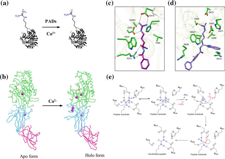
Fig. 1.
a PAD-catalyzed hydrolysis of peptidyl arginine to peptidyl citrulline. b Backbone conformation of PAD2 showing both the apo and holo forms that is generated upon calcium binding. The structural change due to calcium binding is clearly evident in the catalytic domain (green), which harbors the catalytic cysteine C647 (shown in red in the catalytic domain). c Crystal structure of PAD4 C654A protomer bound to the substrate BAA (PDB code 1WDA). d Co-crystal structure of BB-F-amidine (5a) bounds to PAD4 (PDB code 5N0 M). e Proposed catalytic mechanism for PAD4

