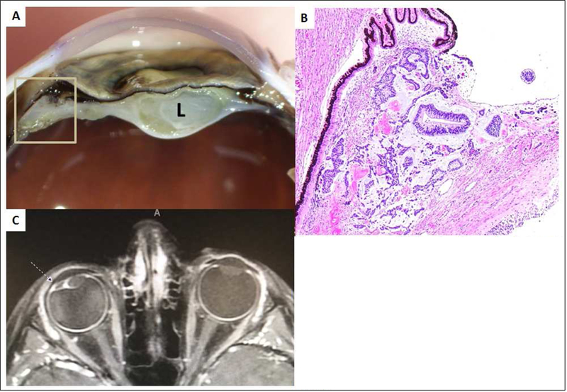
Figure 1.

7-year-old boy, DICER1 carrier with medulloepithelioma of the ciliary body. A: Macroscopic photograph of the anterior portion of the eye after sectioning demonstrating a tan-white lesion (inside square) adjacent to the ciliary body with a membrane extending around the cataractous lens (L). B: Microscopic photograph of the tumor (same site as in the previous photo inside the square) adjacent to the pigmented epithelium of ciliary body. The tumor is composed by tubular structures of neoplastic neuroepithelium seen on a basophilic loose stroma. Hematoxylin and Eosin stain. Original magnification 4X. C: T1 weighted axial image with contrast demonstrating the ciliary body tumor (dashed arrow). Note the displacement of the lens laterally.
