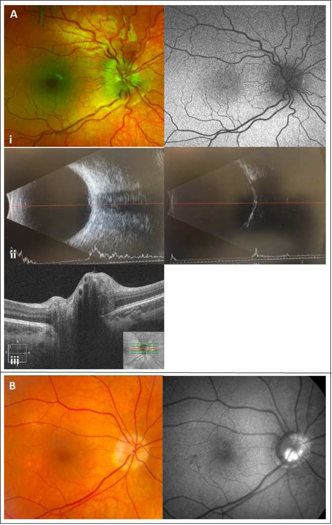
Figure 2.

Panel A: 7-year-old boy with headaches and transient blurred vision. Examination of the fundus revealed disc elevation with retinal vascular tortuosity and no hyperautofluorecence at the nerve head (i). There was no obvious hyper- or hypo-reflective lesion in the peripapillary area on b-scan imaging in high (ii left) or low gain (ii right). OCT does not demonstrate any classic optic nerve head drusen features (iii). Panel B: 65-year-oid female with optic nerve head drusen noted on direct visualization and hyperautofluorescent lesions on imaging.
