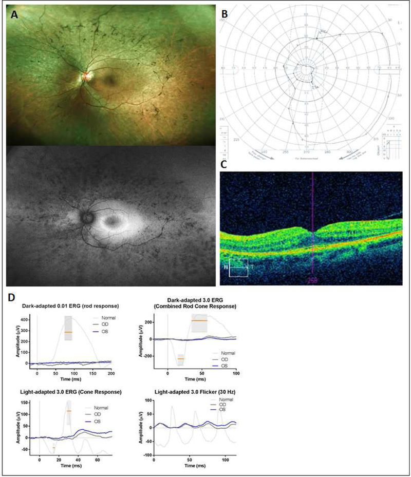
Figure 3.

37-year-old DICER1-carrier woman presenting with retinitis pigmentosa. Fundus examination revealed classic features of retinal atrophy, bony spicules and vessel attenuation (top panel A) with peripheral hypoautofluorescence with a central hyperautoflu orescent ring (lower panel A). Visual acuity was preserved at 20/20 in each eye with a constricted visual field (B) and macular cystic changes and loss of photoreceptor IS/OS band demonstrated on OCT (C). Full-field ERG showed nearly unrecordable scotopic responses with severely reduced photopic responses, consistent with a rod-cone dystrophy (D).
