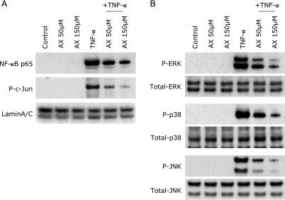
Fig. 6.
Immunoblot analyses for evaluation of NF-κB, AP-1 and MAPK activation in HT-29 colonic epithelial cells. HT-29 cells were stimulated with TNF-α (100 ng/ml) in the presence or absence of astaxanthin for 10 min. Then, the nuclear and cytoplasmic proteins were extracted. (A) Accumulation of NF-κB p65 and phosphorylated (P) c-Jun in the nucleus were evaluated by immunoblotting. (B) The cytoplasmic proteins were subjected to immunoblotting to evaluate phosphorylated ERK1/2, p38 and JNK1. The picture is representative of three independent experiments. AX, astaxanthin.

