Figure 1.

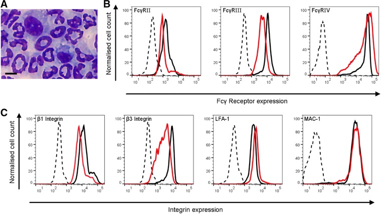
HoxB8 neutrophils express neutrophil integrins and FcγRs. (A) A representative cytocentrifuge preparations of Quick‐Diff stained HoxB8 neutrophils after 5 d of differentiation. Scale bar, 5 μm. (B, C) HoxB8 neutrophil surface FcγRs (B) and integrins (C) as indicated were analyzed by flow cytometry. Representative FACS plots are shown from a minimum of 3 separate experiments performed with HoxB8 neutrophils that had been enriched by separation over a discontinuous percoll gradient. Black traces, BMNs; red/grey traces, HoxB8 neutrophils; broken lines, isotype controls; for FcγRIV, broken line represents secondary antibody only control
