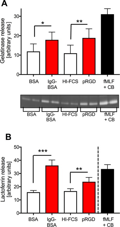
Figure 4.

HoxB8 neutrophils degranulate upon integrin/FcγR stimulation. HoxB8 neutrophils were stimulated by being plated onto pRGD or immobilized ICs, or with 1 μM fMLF in the presence of 10 μM cytochalasin B for 1 h. Released gelatinase (A) and lactoferrin (B) in the supernatant was determined by in gel‐zymography (A) and ELISA (B). A broken line in (B) indicates that the readings obtained with fMLF and cytochalasin B were obtained with more dilute supernatants. Bars show mean ± sem from at least 3 separate experiments performed with HoxB8 neutrophils obtained from 2 different bone marrow donors. *P < 0.05; **P < 0.01; ***P < 0.001; statistical analysis was by paired t test (A) and by unpaired t test (B)
