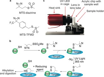
Figure 1.

a) Structures of MTS‐diazirine and MTS‐TFMD. b) Crosslinking workflow schematic: A Cys‐containing bait protein is conjugated with the reagent (here MTS‐diazirine). After adding the target protein, the sample is irradiated with 365 nm UV light, revealing a carbene that reacts with the target. Reductant is added, leaving a sulfhydryl tag on the target at the interaction site. c) Image of the UV LED lamp and custom‐built acrylic chip comprising a 33 μL sample well. See also Figure S4a,b.
