Figure 3.

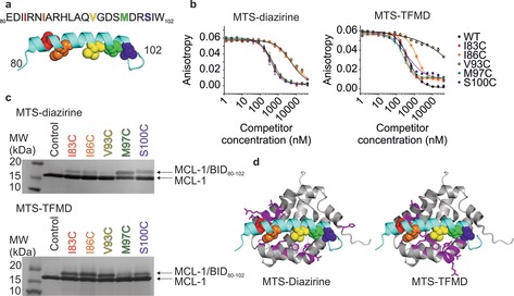
a) Sequence of WT BID80–102. Five Cys variants of BID80‐102 were labelled with MTS‐diazirine or MTS‐TFMD. Cys‐substituted amino acids are shown as coloured spheres in the peptide structure and in the same colour in the sequence above. b) Inhibitory potency (EC50) of BID80–102 labelled with MTS‐diazirine (left) or MTS‐TFMD (right) to MCL‐1 measured by fluorescence anisotropy (EC50 values are shown in Table S1). (c) SDS‐PAGE of BID80–102 labelled with MTS‐diazirine (top) or MTS‐TFMD (bottom) crosslinked to MCL‐1. d) Residues of MCL‐1 (magenta on a grey ribbon) that crosslinked to at least one BID80–102 peptide labelled with MTS‐diazirine (left) or MTS‐TFMD (right). BID80–102 is shown in cyan and residues substituted as Cys are coloured as in (a) (PDB ID: 2KBW16). See also Figures S6–S8, Tables S2, S3.
