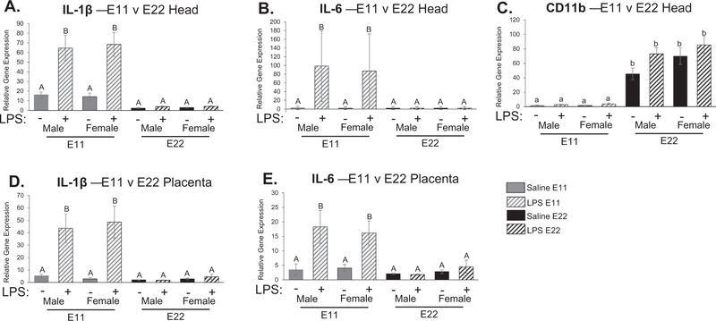
Fig. 5.
Examination of cytokine expression in fetal head and placenta at embryonic day 11 (E11) and embryonic day 22 (E22). There was a significant treatment by condition interaction for IL-1β expression (F1,46 = 36.067; p < 0.001; A) as well as IL-6 expression (F1,45 = 18.225; p < 0.001; B) where gene expression was significantly higher in E11 pups compared to saline treated controls but also compared to E22 pups following LPS administration (p < 0.05). There was a main effect of condition in CD11b gene expression where E22 pups showed significantly higher gene expression compared to E11 pups (F1,46 = 131.846 p < 0.001; C). Similar to fetal brain, there was a significant treatment by condition interaction for IL-1β gene expression (F1,46 = 25.705; p < 0.001; D) and IL-6 expression (F1,46 = 13.472; p = 0.001; E), where inflammatory gene expression was significantly higher in placentas collected at E11 compared to placentas taken at E22 following maternal LPS administration. There were no significant differences between the sexes in the immune response seen in the placenta. *p < 0.05. n = 5–8 rats per group. Condition × Treatment interaction: groups that do not share any capital letters are considered significantly significant (p < 0.05). Main effect of Condition: groups that do not share any lowercase letters are considered statistically significant (p < 0.05).

