
Figure 1.
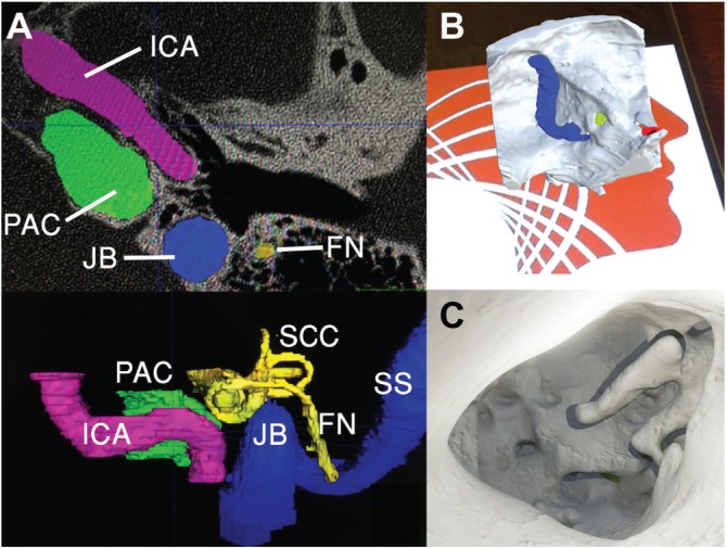
Left ear 3-dimensional (3D) reconstruction. (A) Manual segmentation from computed tomography images into 3D meshes using ITK-SNAP. (B, C) Augmented reality mobile phone application visualized anatomy preoperatively, registered with target image. FN, facial nerve; ICA, internal carotid artery; JB, jugular bulb; PAC, petrous apex cyst; SCC, semicircular canal; SS, sigmoid sinus.