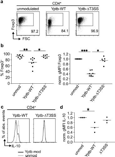
Figure 2.

Foxp3 and IL-10 expression are suppressed in Tregs by Yptb in a T3SS-dependent manner. Foxp3+ Tregs isolated from Foxp3hCD2 reporter mice were co-cultured with Yptb-WT-Bla or Yptb-ΔT3SS-Bla at 37 °C for 1 h at MOI 50. Subsequently, bacteria were killed by antibiotics treatment, and Tregs were cultured on anti-CD3/CD28-coated plates in the presence of IL-2. At day 3 of culture, cells were analyzed by flow cytometry. (a) Representative dot plots show Foxp3 expression among alive CD4+ cells from indicated cultures. Numbers indicate the frequency of Foxp3+ cells in the corresponding gates. (b) Scatterplots summarize the frequencies of Foxp3+ cells (left) and gMFI of Foxp3 (normalized to unmodulated control, right) among alive CD4+ cells from the indicated cultures. (c) Representative histograms show IL-10 expression among alive CD4+ cells from the indicated cultures. (d) Scatterplot summarizes gMFI of IL-10 among alive CD4+ cells from indicated cultures. Each dot represents mean of 3 technical replicates. Data were pooled from 3 to 8 independent experiments (*p < 0.05; **p < 0.01; ***p < 0.001; gMFI, geometric mean fluorescence intensity; MOI, multiplicity of infection; mod, modulated; unmod, unmodulated; T3SS, Type III secretion system)
