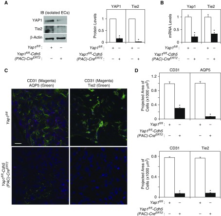
Figure 4.
Knockdown of endothelial YAP1 inhibits vascular and epithelial morphogenesis. (A) Immunoblots (IB) showing YAP1, Tie2, and β-actin protein levels in ECs isolated from Yap1fl/fl-Cdh5(PAC)-CreERT2 or control Yap1fl/fl mouse lungs treated with 4-hydroxytamoxifen (4-OHT) for 48 hours (left). Graph showing the quantification of IB (right, n = 4, *P < 0.05). Readers may view the uncut gels for Figure 4A in the data supplement. (B) Graph showing Yap1 and Tie2 mRNA levels (n = 4, *P < 0.05) in ECs isolated from Yap1fl/fl-Cdh5(PAC)-CreERT2 or Yap1fl/fl mouse lungs treated with 4-OHT for 48 hours. (C) Immunofluorescence micrographs showing CD31-positive blood vessels and AQP5-positive alveolar type I epithelial cells (left) and CD31-positive blood vessels and Tie2 expression (right) in the fibrin gel implanted on Yap1fl/fl-Cdh5(PAC)-CreERT2 or control Yap1fl/fl mouse lungs for 7 days (scale bar: 50 μm). (D) Graphs showing quantification of CD31-, AQP5-, or Tie2-positive cells in the gel implanted on the Yap1fl/fl-Cdh5(PAC)-CreERT2 or Yap1fl/fl mouse lungs for 7 days (n = 7, mean ± SEM, *P < 0.05).

