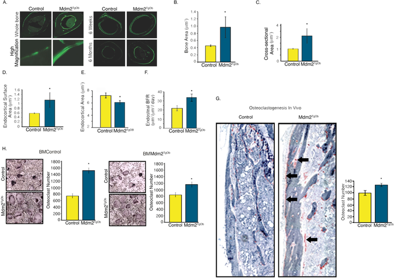
Figure 5. Conditional overexpression of Mdm2 in osteogenic cells increases cortical bone mass and formation.

A. (Left panel) Calcein labeled, femoral midshaft, cross-sections from wild type controls and Mdm2TgOb mice and (Right panel) at 6 weeks and 6 months. B. Bone area. C. Cross-sectional area. D. Endocortical surface area. E. Endocortical area. F. Histomorphometry shows an increase in endosteal bone formation rate (BFR) of Mdm2TgOb compared to wild type controls. G. Femoral TRAP staining (black arrows) and quantitation demonstrating increased osteoclastogenesis in Mdm2TgOb mice. H. In vitro TRAP staining and quantitation using wild type osteoblast progenitors and co-cultured with wild type or Mdm2tgob increased osteoclast (left) or Mdm2tgob osteoblast progenitors cells with bone marrow cells from wild type or Mdm2tgob (right). Asterisk signifies statistical significance at p<0.05.
