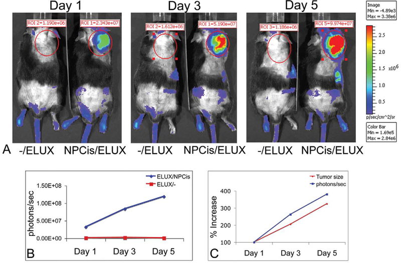
FIGURE 3.—
NPcis/ELUX spontaneous MPNST bioluminescence (A). The bioluminescence (photons/sec) in the NPcis/ELUX tumor is 84-fold higher than background by the end of the experiment (Day 5) (B). The increase in tumor bioluminescence correlates with the increase in tumor size over time (C). Day 1 is the first day on which early tumors can be palpated. Tumor growth in this model is highly variable, but serial imaging allows accurate measurement of tumor growth over time. Representative data is shown for one mouse.

