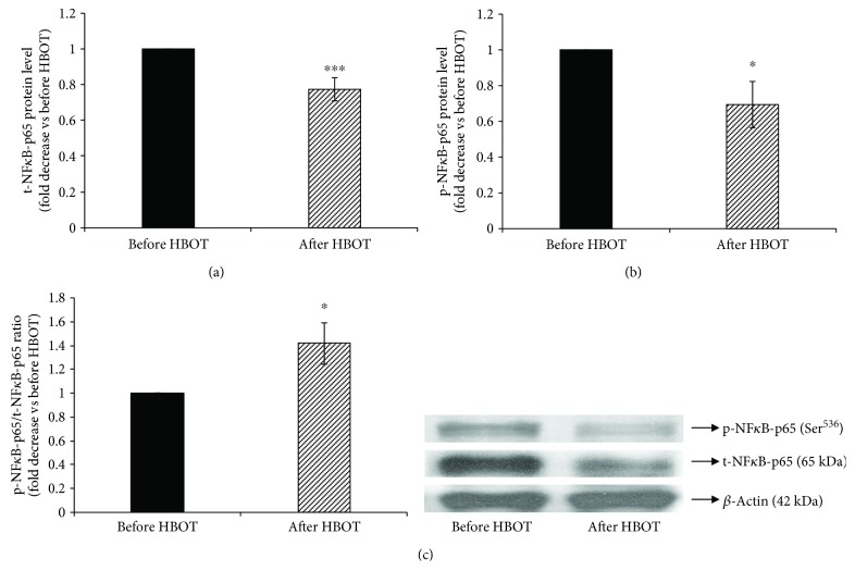
Figure 3.
Phosphorylation of NFκB-p65 at Ser536 in lymphocytes after HBO exposure. (a) Level of t-NFκB-p65 protein in lymphocytes, normalized to β-actin and expressed as fold change before HBOT (arbitrary control set at 1); (b) level of p-NFκB-p65 (Ser536) protein in lymphocytes, normalized to β-actin and expressed as fold change before HBOT (arbitrary control set at 1); (c) ratio of p-NFκB-p65/t-NFκB-p65 expressed as fold change before HBOT (arbitrary control set at 1). Inset: representative Western blots (n = 5–15); ∗∗p < 0.01; ∗p < 0.05; p-NFκB-p65: the phosphorylated form of p65 subunit of nuclear factor-κB; t-NFκB-p65: the total form p65 subunit of nuclear factor-κB. Other abbreviations are under Table 1.

