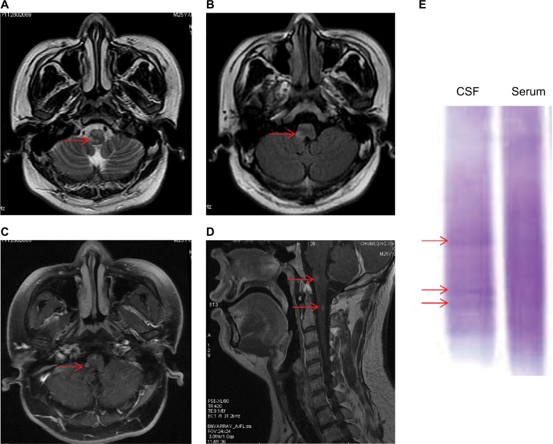
Figure 1.
MRI findings and OB found in the CSF on admission.
Notes: MRI of the brain revealed nodular regions of abnormal signal in the medulla oblongata and cervical cord, characterized by hyper-intensity on (A) T2-weighted and (B) flair sequences (arrow). (C and D) Nodular enhancing lesions were detected in the medulla oblongata and cervical cord (arrow). (E) OB found in the CSF (arrow).
Abbreviations: CSF, cerebrospinal fluid; MRI, magnetic resonance imaging; OB, oligoclonal band.

