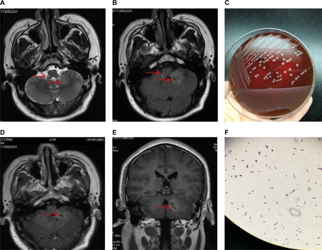
Figure 2.
MRI findings and bacterial cultures when the patient’s condition worsened.
Notes: Repeat MRI was performed and the lesions were found not only in the brainstem but also in the cerebellum with a hyper-intensity on (A) T2-weighted and (B) flair sequences (arrow). (C) Bacterial cultures were taken and Lm was identified. (D and E) The lesions were found patched or ring gadolinium-enhanced with abscess-like appearances (arrow). (F) Gram staining of the bacteria.
Abbreviations: Lm, Listeria monocytogenes; MRI, magnetic resonance imaging.

