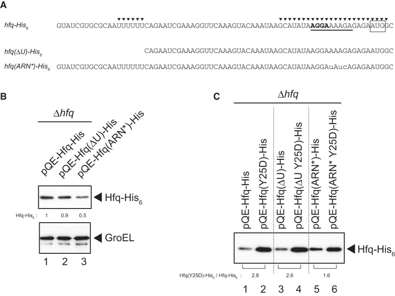
FIGURE 3.
Effects of mutations in 5′-UTR on hfq-His6 expression. (A) RNA sequences of 5′-UTR of the wild-type and mutated hfq genes. The SD sequence is represented by bold letters while the initiation codon is boxed. The two Hfq-binding sites identified previously (Vecerek et al. 2005) are indicated by closed triangles. The sequence corresponding to the (ARN)3 repeat is underlined. The internal U-stretch along with the upstream sequence is deleted in the ΔU mutant. Three nucleotides corresponding to the (ARN)3 are mutated in the ARN* mutant as shown by small letters. (B) Overnight cultures of TM589 (Δhfq) cells harboring indicated plasmids were inoculated (1/100-fold) into LB medium containing 1 mM IPTG. Incubation was continued for 90 min and then total proteins were prepared. Protein samples equivalent to 0.0125 or 0.0025 A600 units were subjected to western blotting using anti-His6 monoclonal antibody or anti-GroEL polyclonal antibodies, respectively. Relative levels of Hfq-His6 derivatives are calculated, with the pQE-Hfq-His sample set to one. (C) Protein samples mentioned above (Fig. 3B) equivalent to 0.0125 A600 units (lanes 1 and 2), 0.0188 A600 units (lanes 3 and 4), and 0.0313 A600 units (lanes 5 and 6) were subjected to western blotting using anti-His6 monoclonal antibody. The relative expression level of Hfq(Y25D)-His6 to Hfq-His6 in each 5′-UTR variant is shown below the gel.

