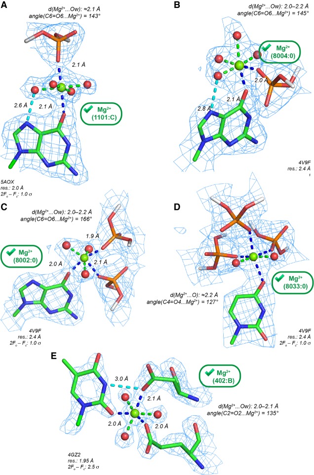
FIGURE 4.
Mg2+ contacting both carbonyl (Ob) and anionic oxygen atoms (Oph, Ocoo) with d(Mg2+…O) ≤ 2.3 Å. The green marks indicate Mg2+ with appropriate densities and stereochemistry. (A) Trans-Oph.Ob: Mg2+ binding to a guanine Hoogsteen edge. A water molecule establishes an H-bond (dashed cyan line) with the (G)N7 atom. (B) Cis-Oph.Ob: Mg2+ binding to a guanine Hoogsteen edge. A water molecule establishes an H-bond (dashed cyan line) with the (G)N7 atom. (C) A ribosomal cis-2Oph.Ob binding site. (D) A ribosomal fac-3Oph.Ob binding site. Note the coordination distances ≈2.2 Å that suggest the use of restraints (Supplemental Table S2). (E) A cis-2Ocoo.Ob binding site at a protein–RNA interface. The contact with a first shell water molecule and a (U)N3-H group, that was considered as difficult to establish (see Fig. 3C), is here replaced by a (U)N3-H…O=C(Asp) H-bond. (C–E) For nomenclature, see Zheng et al. (2015); Ocoo corresponds to oxygen atoms of carboxylate groups.

