Figure 1.
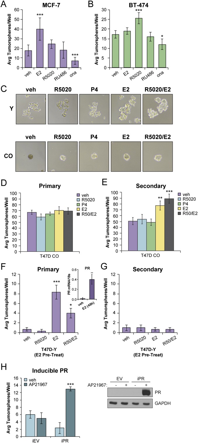
PR expression promotes tumorsphere formation in breast cancer cells. (A) Primary tumorsphere assays in MCF-7 cells. (B) Primary tumorsphere assays in BT-474 cells. Cells were treated with vehicle (veh; EtOH), R5020 (10 nM), E2 (1 nM), RU486 (100 nM), or onapristone (ona; 100 nM). (C) Tumorsphere assays in T47D-Y (PR-null) and T47D CO cells treated with vehicle (EtOH) or the indicated hormone treatment (R5020, 10 nM; P4, 10 nM; or E2, 1 nM). Representative images shown. (D) Primary and (E) secondary tumorspheres in T47D CO cells. (F) Primary and (G) secondary tumorsphere assays in T47D-Y cells. T47D-Y cells were pretreated with E2 (1 nM) for 48 hours, followed by tumorsphere assays with vehicle (EtOH) or the indicated hormone treatment. (F, Right Inset) mRNA levels of PR from primary tumorspheres in T47D-Y cells pretreated with E2. (H) Secondary tumorsphere assays in T47D-inducible EV control and PR-B cells. PR expression was induced within 24 hours after addition of AP21967 (1 nM); Right, Western blot. Graphed data represent the mean ± SD (n = 3). *P < 0.05; **P < 0.01; ***P < 0.001.

