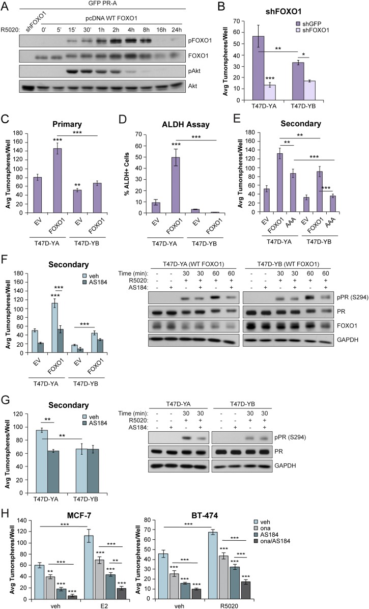
Figure 6.
FOXO1 promoted breast CSC outgrowth in PR isoforms. (A) Western blot of FOXO1 (phosphorylated and total) and Akt (phosphorylated and total) in HeLa cells transfected with green fluorescent protein–tagged PR-A and with pcDNA FOXO1. Cells were treated with vehicle (veh; EtOH) or R5020 (10 nM) for the indicated times. shFOXO1 was included as a control. (B) Primary tumorsphere assays in T47D-YA and T47D-YB cells with FOXO1 knockdown. (C) Primary tumorsphere assays in T47D (YA and YB) cells stably expressing FOXO1. (D) ALDH activity in primary tumorspheres formed from T47D (YA and YB) FOXO1+ cells was assessed by flow cytometry. (E) Secondary tumorspheres in T47D (YA and YB) cells expressing WT FOXO1 or FOXO1-AAA. (F) Secondary tumorspheres in T47D (YA and YB) FOXO1+ cells treated with AS1842856 (100 nM). (Right) T47D (YA and YB) cells expressing FOXO1 were pretreated with AS1842856 (100 nM) for 60 minutes, followed by the indicated treatments with R5020 (10 nM). (G) Secondary tumorspheres in T47D (YA and YB) cells treated with AS1842856. (Right) T47D (YA and YB) cells were pretreated with AS1842856 (100 nM) for 60 minutes, followed by treatment with R5020 (10 nM). (H) MCF-7 and BT-474 primary tumorspheres treated with E2 (1 nM) or R5020 (10 nM), respectively, in the presence of onapristone (100 nM) or AS1842856 (100 nM), or both. Graphed data represent the mean ± SD (n = 3). *P < 0.05; **P < 0.01; ***P < 0.001. Avg, average.

