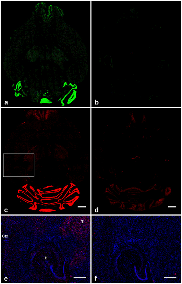
Fig. 1.
Overall distribution and specificity of fluorescein-tagged (a, b), and digoxigenin-tagged (c-f) antisense RNA probe binding in horizontal mouse brain sections. (a, c) GluN2C mRNA signal in wild-type mouse brain showing the highest expression levels in the granular layer of the cerebellum. b, d, GluN2C-KO mouse brain sections showing very low (b) and low (d) background staining in cerebellum and olfactory bulb that was independent of the tag used. (e, f) Enlarged images of the cortex, hippocampus, and thalamus which are highlighted by the box in (c). Scattered GluN2C signal is displayed in these regions of wild-type mouse (e) but not in GluN2C-KO mouse (f). T, thalamus; Ctx, cortex; H, hippocampus. Scale bars: c, d, 1 mm; e, f, 500 μm

