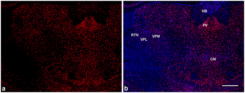
Fig. 7.
GluN2D mRNA distribution in the thalamus in coronal brain sections. (a, b) GluN2D signal (red) is higher in the midline thalamic nuclei (including paraventricular (PV) and central medial (CM) nuclei) compared to the habenula (HB) and the lateral thalamus - ventrobasal complex (VPM/VPL) and RTN. (b) GluN2D mRNA probe (red) co-stained with DAPI (blue). Scale bar = 500μm

