Figure 1.
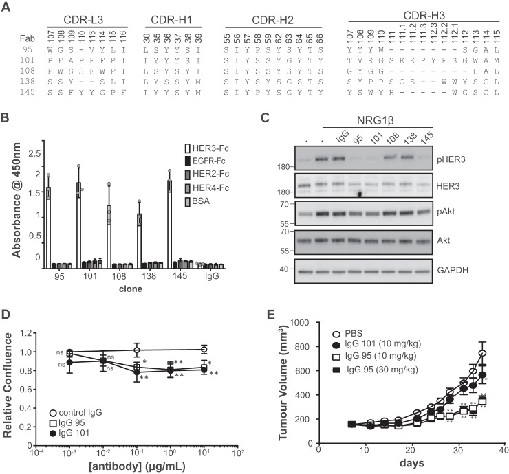
Characterization of anti-HER3 antibodies. A, sequences of anti-HER3 Fabs. Sequences of CDR positions randomized in the library are shown and are numbered according to the IMGT standards (72). B, IgG specificity for ErbB family members assessed by ELISA. Error bars represent the standard deviation of three independent experiments, and each point the mean of one experiment. C, Western blots of lysates from SKBR3 cells treated with the indicated IgG (5 μg/ml) for 1 h prior to treatment with NRG1 (2 nm) for 10 min. Blots were developed with the antibodies to the proteins indicated on the right. The data are representative of three independent experiments. D, cell proliferation assays with SKBR3 cells treated with the indicated antibodies. Relative confluence was measured after control cells doubled 1.5 times (∼55 h). Error bars represent the standard deviation of three independent experiments. *, p < 0.05; **, p < 0.005, one-way ANOVA (see “Experimental procedures”). E, in vivo xenograft assays. Subcutaneous BxPC3 xenografts were established in CB-17 SCID mice and treated with the indicated antibodies. Tumor size was measured at the indicated time points (n = 10 for the PBS control, and n = 9 for all other treatment groups). **, p < 0.0001; *, p = 0.0001, two-way ANOVA.

