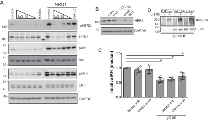
Figure 3.
Effects of IgG 95 on HER3 internalization and ubiquitination. A, Western blots of lysates from SKBR3 cells treated with the indicated IgGs for 90 min. The following concentrations were used for IgG 95: 0.04, 0.2, 1, or 5 μg/ml, and 5 μg/ml was used for IgG control and trastuzumab. The indicated cells were stimulated with 2 nm NRG1 for 10 min before harvest. Blots were developed with the antibodies indicated to the right. The data are representative of 3 independent experiments. B, Western blots of lysates from SKBR3 cells treated with 100 μm chloroquine (chlor) or 50 nm bortezomib (bort) for 1 h prior to treatment with 1 μg/ml of IgG 95 for 2 h, as indicated. Blots were developed with antibodies to the proteins indicated to the right. Representative of three independent experiments. C, internalization of IgG 95. SKBR3 cells were treated with 100 μm chloroquine or 50 nm bortezomib for 1 h prior to treatment with 1 μg/ml of IgG 95 for 2 h, as indicated. The cells were then processed for flow cytometry by staining with IgG 95. Error bars represent the S.D. of 4–5 independent experiments, and each point the relative median fluorescent intensity of a single experiment. *, p < 0.005, one-way ANOVA. D, Western blots for assessment of ubiquitination of HER3. The indicated cells were treated with 100 μm chloroquine for 30 min followed by treatment with 1 μg/ml of IgG 95 for 30 min, as indicated. Lysates were subjected to immunoprecipitation with IgG 95, followed by development with antibodies to the proteins indicated to the right. The data are representative of three independent experiments.

