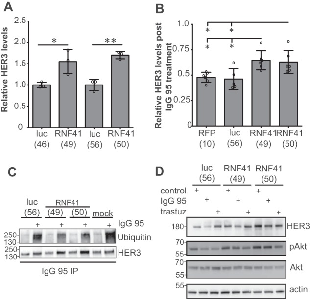
Figure 5.

RNF41 depletion protects HER3 protein from degradation induced by IgG 95. A, quantification by Western blotting of HER3 in lysates from SKBR3 cells stably expressing the indicated shRNA. Error bars represent the S.D. of three independent experiments, and each point the value of one experiment. *, p < 0.05; **, p < 0.005, t test. B, quantification by Western blotting of HER3 in lysates from SKBR3 cells stably expressing the indicated shRNA and treated with 1 μg/ml of IgG 95 or control IgG for 2.5 h. Error bars represent the S.D. of at least five independent experiments, and each point the value of one experiment. *, p < 0.05, one-way ANOVA. C, Western blots of IgG 95 immunoprecipitates from SKBR3 cells stably expressing the indicated shRNA and pretreated with 100 μm chloroquine for 30 min, and then with 1 μg/ml of control or IgG 95 for 30 min. Representative data from one of three independent experiments are shown. D, Western blots of lysates from SKBR3 cells stably expressing the indicated shRNA and treated with 5 μg/ml of the indicated IgG for 2.5 h. The data are representative of three independent experiments.
