Figure 1.
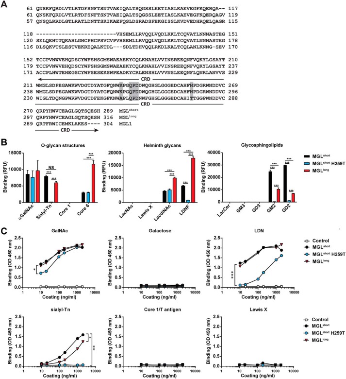
Differential GalNAc binding of MGL variants. A, amino acid sequence of the human MGL splice variants and the mouse MGL1. The stalk region and CRD are depicted, showing the 27-amino acid insertion in the stalk region of the MGLlong variant. Boxed in gray are the primary binding site (QPD) of MGL and the localization of the K237A and H259T mutations. B, binding of MGL variants determined by glycan array. Depicted are the binding to selected O-glycan core structures (left), Helminth-associated glycans (middle), and glycosphingolipids (right). Full glycan array results can be found in Table S1. Significance was calculated using two-way analysis of variance and Bonferroni's multiple-comparison test. Data represent mean ± S.E. LacCer, lactosylceramide. C, binding of the MGL variants to PAA-coated glycoconjugates was determined using an ELISA-based assay. Human IgG1 was used as a negative control. One representative experiment of three is shown. Significance was calculated by nonlinear regression followed by one-way analysis of variance and Bonferroni's multiple-comparison test. *, p < 0.05; **, p < 0.01; ***, p < 0.005.

