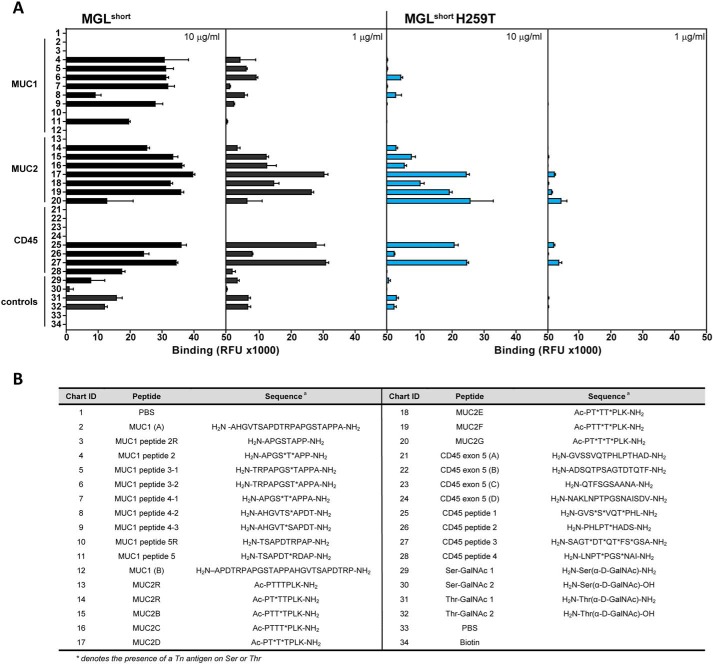
Figure 2.
Binding profile of MGL variants to the Tn-glycopeptide array. A, glycopeptides and corresponding parental peptides were printed at 100 μm and probed using the MGLshort-Fc (left panels) and MGLshort H259T-Fc (right panels) at the indicated concentrations. Relevant protein carriers are indicated in the figure. Data represent mean ± S.D. (error bars). RFU, relative fluorescence units. B, list of peptide backbones and Tn attachment sites of glycopeptides present on the Tn-glycopeptide array. *, presence of a Tn antigen on Ser or Thr.

