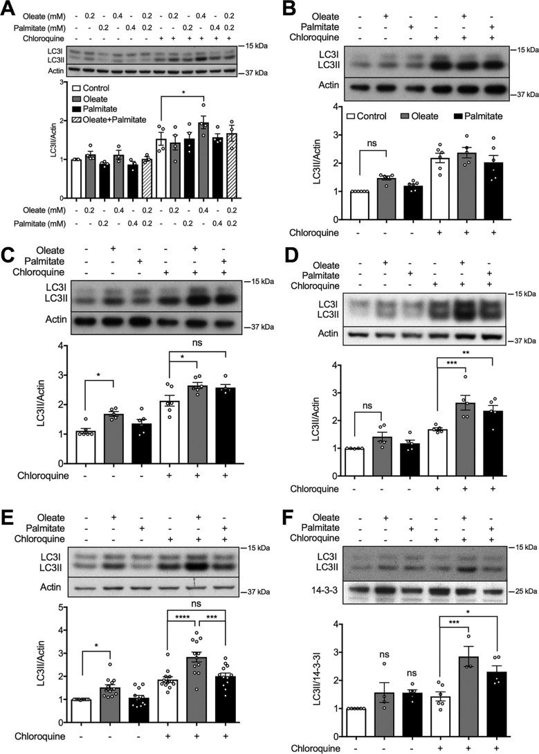
Figure 1.
Effect of unsaturated and saturated FAs on autophagic flux in MIN6 cells or mouse islets. A–E, levels of the autophagic marker LC3II in MIN6 cells after treatment with (A) oleate or palmitate, alone or combined, at the indicated concentrations for 48 h; or 0.4 mm oleate or 0.4 mm palmitate for (B) 8, (C) 24, or (D and E) 48 h (n = 5–12). F, levels of LC3II in isolated mouse islets with 48 h treatment of 0.4 mm oleate or 0.4 mm palmitate (n = 4). The BSA-treated group was used as the control. Where indicated MIN6 cells or islets were incubated with or without 50 μm CQ for 2 h prior to cell lysis. Data shown are the mean ± S.E. of the densitometric quantification. Statistical analyses were done with one-way ANOVA with Sidak post hoc test. *, p < 0.05; **, p < 0.01; ***, p < 0.001; and ****, p < 0.0001.

