Figure 2.
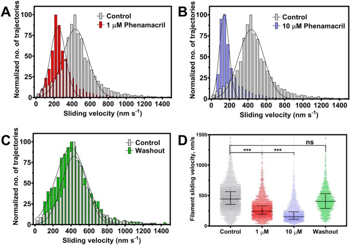
Functional inhibition of FgMyo1IQ2 by phenamacril. A–C, trajectory-normalized distribution of the pooled sliding velocities of rhodamine-phalloidin–labeled F-actin filaments on a lawn of FgMyo1IQ2 before and after the infusion of phenamacril. The average sliding velocity decreases with the sequential infusion of 1–10 μm phenamacril (A and B) and is restored upon inhibitor washout (C). D, scatter plot of filament sliding velocities with median and interquartile range from three independent experiments. *** and ns denote that the differences between experiments were significant (p < 0.0005) or not significant, respectively.

