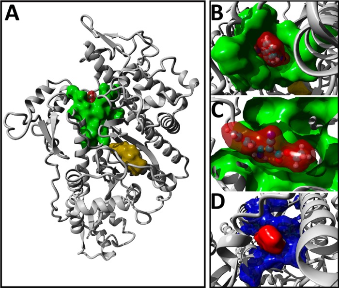
Figure 5.
Blind-docking of phenamacril is consistent with a noncompetitive mode of inhibition. A, the position of the top-ranking docking pose of phenamacril (red surface-rendering) in FgMyo1 is shown, highlighting contact residues (green). For reference, the position of ADP-vanadate (yellow) was superimposed into the model. B and C, top view (B) and cut-plane side view (C) of phenamacril and the molecular surface of amino acid residues involved in the FgMyo1-phenamacril interaction. D, surface-rendering of the phenamacril docking-pose (red) and position of residues (blue), which are known to mutate and confer resistance to phenamacril.

