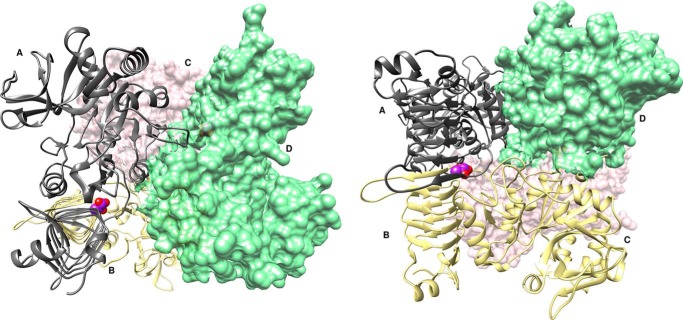
Figure 1.
Complex structure of the A. tumefaciens ADP–Glc PPase (mutant P96A) bound to pyruvate. The ribbon and surface structure represent a homotetramer bound to pyruvate (PDB code 5W5R). The enzyme can also be referred to as a dimer of dimers with subunits A (gray ribbon) and B (yellow ribbon) forming one dimer and subunits C (pink surface) and D (green surface) forming the other. Each dimer binds one pyruvate molecule, shown as purple spheres, and heteroatom oxygen atoms, shown as red spheres, with a stoichiometry of two pyruvate molecules per tetramer.

