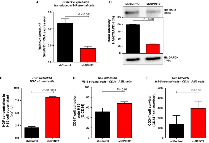
Figure 2.

SPINT2 silencing induces HGF secretion, hematopoietic cells adhesion onto HS‐5 stromal cells and CD34+ cells survival. (A) Quantitative expression of SPINT2 mRNA in shSPINT2 cells relative to the shControl cells in HS‐5 stromal cells. Lentivirus‐mediated SPINT2 shRNA effectively silenced SPINT2 in HS‐5 stromal cells. mRNA expression levels of SPINT2 were normalized by HPRT endogenous control, as indicated. Results were analysed using 2−ΔΔ CT. Experiments were performed in triplicate. P values are indicated. (B) Western blot analysis of shControl and shSPINT2 total cell extracts of HS‐5 stromal cells. The membrane was blotted with antibodies against SPINT2/HAI‐2 (34 kDa) or actin (42 kDa), as a control for equal sample loading, and developed with the SuperSignal West Pico Chemiluminescent Substrate (Thermo Scientific). (C) Analysis of HGF secreted by shControl and shSPINT2 HS‐5 after 48 hours of culture. (D) CD34+ cells from de novo AML patients were added to a monolayer of nontransduced and shControl and shSPINT2 HS‐5 stromal cells and allowed to adhere for 24 hours. After 24 hours, nonadherent cells were removed by gentle aspiration, and the CD34+ cells that adhered to HS‐5 stromal cells were collected by gentle pipetting cold phosphate‐buffered saline. The percentage of CD34+ adherent cells were measured by flow cytometry using CD34‐APC and analysed as percentage of total cells. Percentages of cells expressing the marker were determined out of a total 10,000 events. (E) CD34+ cells from de novo AML total bone marrow were added to a monolayer of nontransduced and shControl and shSPINT2 stromal cells and cultured for 48 hours. After 48 hours, nonadherent cells were carefully collected by gentle aspiration, labelled with CD34‐APC antibodies and measured by flow cytometry. Values are means ± standard deviation of three independent experiments. Statistical analysis: Mann‐Whitney test. P values are indicated
