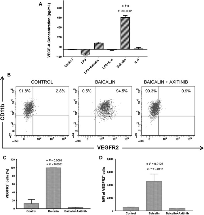
Figure 3.

Autocrine VEGF milieu of M2 macrophage interfered by VEGF receptor inhibitor. A, The secreted VEGF‐A protein in conditioned media were measured. B, C, The percentage of M2 macrophages expressing VEGF‐R2. D, The mean fluorescence intensity (MFI) of VEGF‐R2 on M2 macrophages. The Data were determined by mean ± SEM, n = 3. *P < 0.05 compared with control group; # P < 0.05 compared with LPS; ‡ P < 0.05 compared with IL‐4; ≠P < 0.05 compared with axitinib. VEGF, vascular endothelial growth factor; LPS, lipopolysaccharide; IL‐4, interleukin‐4
