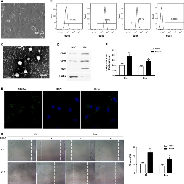
Figure 1.

Mesenchymal stem cells (MSC)‐derived exosomes reduced proliferation and migration of vascular smooth muscle cells (VSMCs). (A) The morphology of MSCs was observed under a microscope; scale bar = 100 μm. (B) Representative fluorescence‐activated cell sorting (FACS) analysis of MSCs expressing antigens for CD90, CD29, CD44 and CD34. C. Micrographs obtained by transmission electron microscopy of MSC‐derived exosomes. (D) Western blot assays to detect exosome/extracellular vesicle‐specific markers (CD63, CD81, CD9) in MSCs and purified exosomes. (E) Representative micrographs obtained by confocal microscopy of DIO‐labelled exosome (green) uptake by VSMCs. Nuclear counterstaining was performed with DAPI (blue). (F) Analysis of VSMC proliferation by MTT assays. VSMCs were treated with or without MSC‐Exo in the presence or absence of PDGF‐BB (20 ng/mL) for 48 h. Three independent experiments (n = 3). (G) Cell migration was measured after treatment with or without MSC‐Exo in the presence or absence of PDGF‐BB (20 ng/mL) over 24 h by scratch‐wound assays and is presented as the percentage of cell closure. Experiments were repeated at least three times (n = 3) and data are the average of repeat experiments. **P < 0.01 vs no PDGF treatment, # P < 0.05 vs control cells
