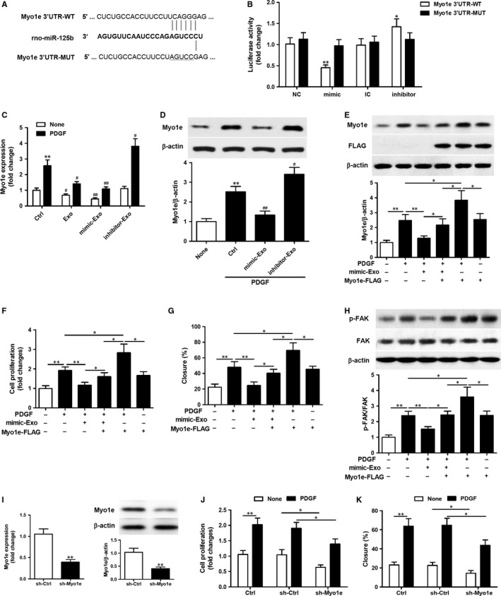
Figure 4.

miR‐125b shuttled by mesenchymal stem cells (MSC)‐exosomes suppresses vascular smooth muscle cells (VSMC) proliferation and migration by targeting Myo1e. A. Schematic representation of the homologous sequence between rat Myo1e 3ʹUTR mRNA and miR‐125b. B. Dual luciferase assay on wild‐type (WT) or mutant (MUT) rat Myo1e 3ʹUTR in VSMCs transfected with miR‐125b mimic (or mimic control, NC) or inhibitor (or inhibitor control, IC). Data represent the luciferase activity ratio of firefly to Renilla luciferase. *P < 0.05, **P < 0.01 vs NC or IC. Three independent experiments (n = 3). (C) RT‐qPCR analysis of the expression of Myo1e in VSMCs after treatment with exosomes isolated from MSCs or MSCs transfected with miR‐125b mimic or inhibitor in the presence or absence of PDGF‐BB (20 ng/mL) for 24 h. **P < 0.01 vs no PDGF‐BB control; # P < 0.05, ## P < 0.01 vs control cells. Three independent experiments (n = 3). (D) Western blot analysis of Myo1e in VSMCs after treatment with exosomes isolated from MSCs transfected with miR‐125b mimic or inhibitor in the presence of PDGF‐BB (20 ng/mL) for 24 h. **P < 0.01 vs no PDGF‐BB control; # P < 0.05, ## P < 0.01 vs control cells. β‐actin was detected as a loading control. Three independent experiments (n = 3). (E‐H) VSMCs transfected with Myo1e overexpression plasmid for 24 h were stimulated with or without exosomes isolated from MSCs transfected with miR‐125b mimic in the presence of PDGF‐BB (20 ng/mL). (E) Western blot analysis of Myo1e expression in the treated VSMCs. (F) Analysis of VSMC proliferation by MTT assays. (G) The percentage of cell closure as measured by scratch‐wound assays. (H) Western blot analysis to examine FAK phosphorylation. β‐actin was detected as a loading control. (I‐K) VSMCs transfected with lentiviral particles of the Myo1e shRNA and lenti control shRNA for 48 h, stimulated with or without PDGF‐BB (20 ng/mL) for 24 h. (I) RT‐qPCR analysis and Western blot analysis of Myo1e expression in the VSMCs. (J) Analysis of VSMC proliferation by MTT assays. (K) The percentage of cell closure as measured by scratch‐wound assays. *P < 0.05, **P < 0.01. Three independent experiments (n = 3)
